
It emerges from the confluence of the superficial submandibular veins beneath the chin and drains into the external jugular vein. Internal jugular vein ectasia dilatation of the internal jugular vein is a rare clinical entity often undiagnosed.
Bilateral Internal Jugular Phlebectasia
Our report describes right internal jugular vein ectasia in a 15-year-old girl who presented to us with intractable paroxysmal cough.

. Internal jugular external jugular or superficial veins are usually the affected ones in the neck. This also was not con-tributory and therefore her parents were informed that the ectatic internal jugular vein was the only abnormality detected. As an ectatic internal jugular vein had never been implicated in the past as a cause of paroxysmal cough and as no other cause could be found the patient and her parents were subjected to psychological evaluation and counselling.
The exact cause of this. Jugular vein ectasia is a venous anomaly that commonly presents itself as a unilateral neck swelling in children and adults. Here we are reporting a patient with venous ectasia of the retromandibular vein and the common facial vein.
We conclude that internal jugular venous ectasia in children is a benign condition which usually does not require surgical intervention. Internal jugular vein ectasia is a venous anomaly commonly presenting as a unilateral neck swelling in children and adults. Internal jugular vein ectasia dilatation of the internal jugular vein is a rare clinical entity often undiagnosed.
Like arterial aneurysms venous ectasias are caused by structural weaknesses of the vessel wallthis could be the result of local trauma inflammation a congenital weakness localized degenerative changes and possibly elevated vascular flow and pressure. This condition is being increasingly recognized in recent years due to better imaging facilities. Ultrasonography is a good diagnostic modality for the diagnosis of internal jugular venous ectasia.
Venous ectasia dilation of veins or venules such as. They are often ignored or misdiagnosed. Venous ectasias are benign conditions of the neck in which focal dilatations of veins occur.
The exact cause of this. Our report describes right internal jugular vein ectasia in a 15-year-old girl who presented to us with intractable paroxysmal cough. It is important to include it in the.
The jugular veins are found in the neck. The external jugular veins empty into the subclavian veins. There is a controversy about its etiology because there have been only sporadic reports of venous ectasia in the neck.
3 2009 Focal Ectasia of Internal Jugular Vein 283 on the right side 3. High index of suspicion is required to diagnose the condition. This condition is being increasingly recognized in recent years due to better imaging facilities.
Telangiectasias are small dilated blood vessels found anywhere on the body but commonly seen on the face around the nose cheeks and chin. Chronic venous insufficiency often in the leg Jugular vein ectasia in the jugular veins returning blood from the head See also. Usually it presents as an asymptomatic soft compressible neck swelling that increases in size on Valsalvas manoeuvre.
These rare lesions appear to be congenital dilatations of the veins and should not properly be called aneurysms as their histological structure is normal. There is a controversy about its etiology because there have been only sporadic reports of venous ectasia in the neck. Neck contrast-enhanced computed tomography revealed a venous dilatation in the right retromandibular vein region near its confluence with the facial vein associated with ectatic ipsilateral external and internal jugular veins Download.
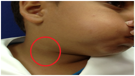
It is commonly seen in children and young adults 1 2 3 and appears as soft compressible mass in the neck which becomes prominent on straining or performing Valsalva maneuver. Bilateral internal jugular vein ectasia. Vascular anomalies can be divided into two groups as vascular tumor and vascular malformation according to ISSVA 4.
Ultrasonography with Doppler before during and after valsalva manoeuvre is the preferred method for diagnosis. Intrinsic vascular malformations arising from the wall of internal jugular vein have not been described in English-language literature. 5 the etiology of retromandibular vein aneurysms are likely similar to those of other.
It is rare to have bilateral neck swelling due to internal jugular vein ectasia. Die Vena jugularis externa formiert sich innerhalb der Ohrspeicheldrüse Glandula parotis in der Höhe des Kieferwinkels aus dem Zusammenfluss der Vena auricularis posterior und der Vena retromandibularis gelegentlich auch aus dem Zusammenfluss der Vena auricularis posterior und der Vena occipitalis. Few cases of intrinsic vascular malformations arising from external jugular vein have been reported 3 4We describe a case of vascular malformation of internal jugular vein which presented in an adult as a slowly enlarging mass at.
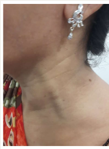
A typical history and complex of physical findings are associated with this entity. A 25-year-old male presented. Jugular vein ectasia is a venous anomaly that commonly presents itself as a unilateral neck swelling in children and adults.
Color Doppler ultrasonography demonstrate the turbulent flow in jugular venous ectasia. They are the main path for deoxygenated blood returning from the cranium back to the heart. The treatment is conservative for asymptomatic patients and surgery is reserved for patients with.
Download high-res image 536KB Download. There is a pair of internal jugular veins right and left and a pair of external jugular veins. Focal saccular aneursymal dilatation of the proximal right internal jugular vein.
It is rare to have bilateral neck swelling due to internal jugular vein ectasia. Three cases of jugular venous ectasia in children were diagnosed preoperatively by selective venography. It can be primary congenital or acquired due to trauma surgery infection and tumors.
Internal jugular vein ectasia is a venous anomaly commonly presenting as a unilateral neck swelling in children and adults. The anterior jugular vein is a paired blood vessel that drains the anterior aspect of the neck. Download full-size image Fig.
Ectasia of external jugular vein is a rare entity presenting as an intermittent neck swelling. The right internal jugular vein valves are placed at a higher level than the left sided ones and. Case Discussion Saccular aneursyms of the internal jugular vein are rare as opposed to fusiform aneursyms phlebectasia found in the pediatric population.
Usually it presents as an asymptomatic soft compressible neck swelling that increases in size on Valsalvas manoeuvre. Jugular vein ectasia is a rare benign condition.

Internal Jugular Phlebectasia In An African Adult Bmj Case Reports
2

Bilateral Internal Jugular Phlebectasia
Unilateral Right Sided Internal Jugular Vein Ectasia In A Woman At Rest Download Scientific Diagram

Ijv Phlebectasia An Approach Algorithm Semantic Scholar

A Boy Who Can Puff His Neck Bilateral Internal Jugular Phlebectasia Pediatrics Neonatology
Medcrave Online

Bilateral Internal Jugular Phlebectasia

Normal Appearance Of Neck At Rest A Unilateral Right Sided Internal Download Scientific Diagram
2

Pin On Shoulder Pain Research

Internal Jugular Phlebectasia A Case Report And Literature Review Semantic Scholar
Left External Jugular Phlebectasia Rare Presentation In Adults Sign Of A Deep Dangerous Lesion Crimsonpublishers Com
2
2

Neck Masses Due To Internal Jugular Vein Phlebectasia Frequency In Menkes Disease And Literature Review Of 85 Pediatric Subjects Stevens 2020 American Journal Of Medical Genetics Part A Wiley Online Library
:max_bytes(150000):strip_icc()/Jvd-causes-5185667_final-eff74efaccdf4040a69177ee6109b38e.jpg)
Jugular Vein Distention Common Causes Of Jvd
2
2